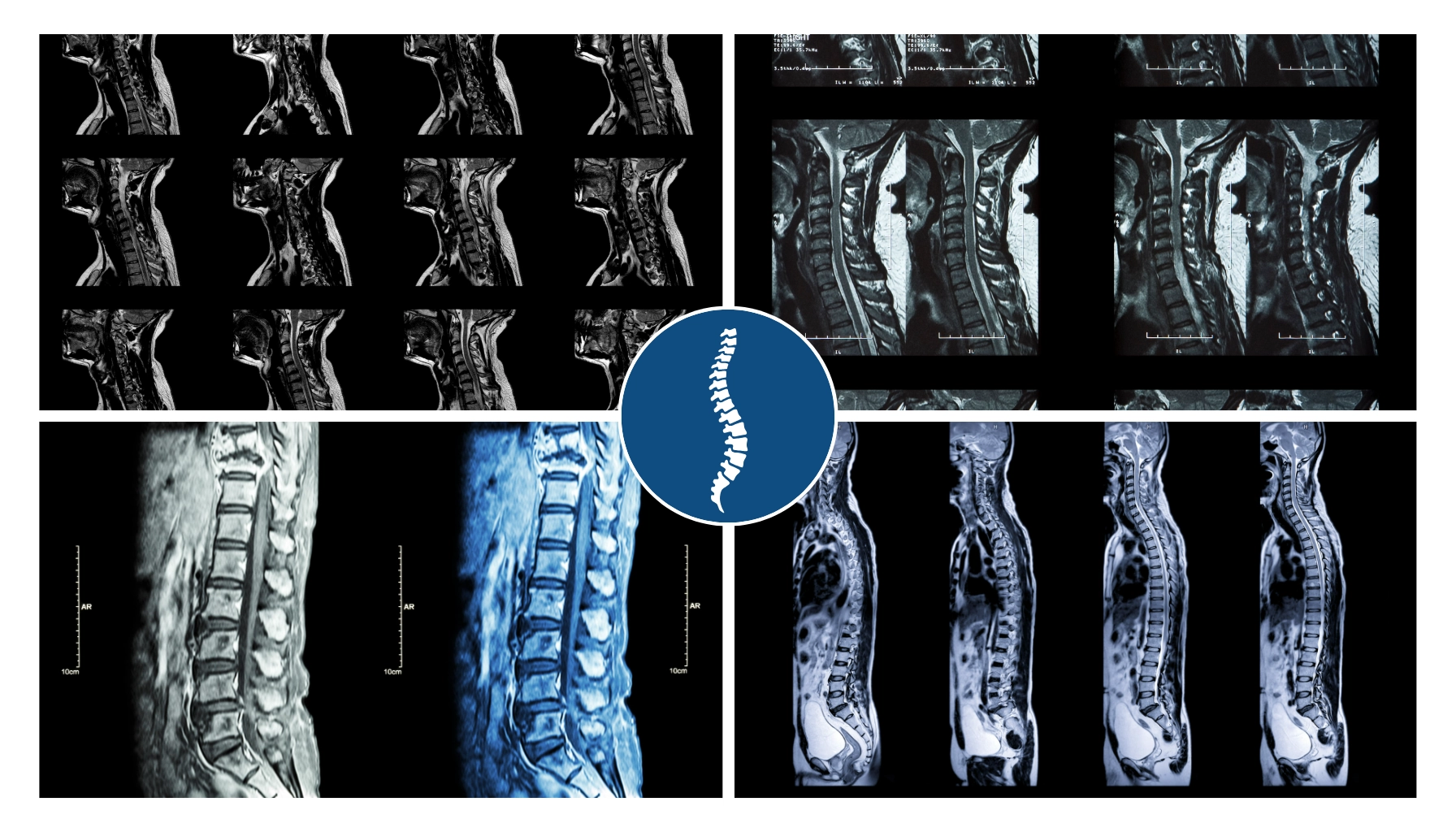
A spine MRI report describes what your spine looks like on the day of the scan, and most of the findings on it also appear in people who have no back pain.
Patients usually arrive with their spine MRI report in hand and a few phrases from it already worrying them. Degeneration, bulge and narrowing all sound serious, and many people have not been told how ordinary those words are. I start by asking where it hurts and for how long, because the scan comes second.
What a spine MRI report actually shows
An MRI shows structure, while pain is something your nervous system produces, and the two disagree more often than most people expect. A review of scans from 3,110 adults with no back pain found disc degeneration in 37% of adults aged 20, and in 96% of those aged 80. The scans were accurate and the findings were real, but none of those people had symptoms, so a finding on its own cannot tell you it is the source of your pain.
Five findings you will see on most reports
- Disc desiccation or water loss
A disc holds water and holds less of it with age, so it looks darker on the scan. It tells me the disc is older, not that it has been hurt.
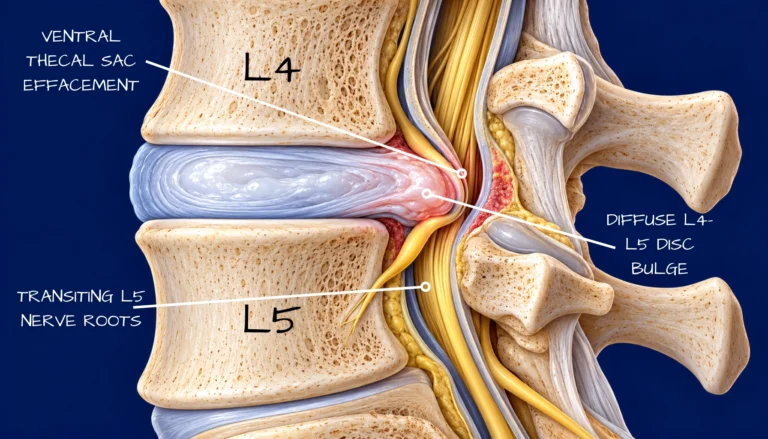
- A disc bulge or protrusion
Disc material sits slightly beyond its normal edge, and half of adults in their forties with no back pain have one. A disc herniation, where the material presses on a nerve, is a different and more specific finding.
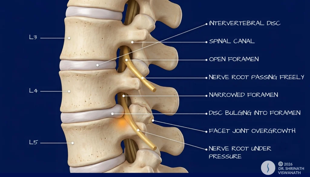
- Neural foraminal narrowing
The foramen is the opening where a nerve root leaves the spine on its way to the arm or leg, and narrowing means less room there. It is common with age and it only matters if the level and side match where you feel symptoms.
- Annular fissure or tear
This is a small crack in the tough outer wall of the disc. It can be a source of local pain and it also turns up in people who have none.
- Loss of lordosis
The normal inward curve of the lower back looks flatter on the images. It often reflects muscle tension or how you were lying in the scanner more than any structural problem.
What your spine MRI report cannot tell you
It cannot tell me whether the pain is coming from the spine at all, how severe it is, or whether you need surgery, physiotherapy or simply time. Two patients can bring me almost identical reports and leave with different plans, because the difference lives in their history and examination. A repeat scan can even show more change while the person feels better, since the scan follows anatomy and your experience follows something else.
What to prepare before you see a spine surgeon
Think about where the pain starts, whether it travels into the leg, and which movements make it better or worse, and notice any change in strength or sensation. Write the answers down beforehand, because they are easy to forget once you are in the room.
When to See a Doctor
Arrange a spine assessment if:
PLAN AN EVALUATION
- Back or leg pain has lasted longer than six to eight weeks without meaningful improvement
- Numbness in the saddle area, meaning the inner thighs, groin or around the anus
- Weakness in one or both legs that is getting worse quickly
These may be signs of cauda equina syndrome, a condition in which the nerve bundle at the base of the spinal cord is compressed. This requires same day orthopaedic or emergency evaluation. Do not wait.
COMMON QUESTIONS
My spine MRI report says severe degeneration. Does that mean I need surgery?
No. Severity on a scan describes how much the structure has changed, not how much it hurts or what you need. If your report mentions a bulge or herniation, my article on what a disc bulge actually means explains the difference.
Q2: Should I avoid exercise because of what the scan shows?
For most people no. Guided movement is usually appropriate even when degeneration is visible on imaging, and avoiding activity because of a scan finding rarely helps. I advise on this after examining you.
Q3: Do I need an MRI before seeing a spine surgeon for the first time?
Not always. A history and examination often tell me more than a scan done before we have met, and I usually order imaging once I know the question it has to answer.
Q4: If my spine MRI report mentions Modic changes, should I be worried?
Modic changes are signal changes in the vertebral bone next to a disc, and they most often reflect wear and tear. Occasionally a similar picture comes from infection, which is why I read them alongside your symptoms and examination.

Pingback: Back Pain: 4 Patterns That Reveal the Real Source