By Dr. Shrinath Viswanath, Orthopedic Spine Surgeon · Last reviewed: 7th July 2026
Overview
A spine MRI report uses technical terms — disc bulge, foraminal narrowing, degenerative changes — that sound alarming but often have straightforward explanations. Most MRI findings are manageable, and many are present in people with no pain at all. Understanding what these terms actually mean helps you have a more informed conversation with your spine specialist and helps you clarify what does your spine MRI report mean.
Almost every week, a patient walks into my consulting room holding a spine MRI report, points to a line they have circled in pen, and asks me: “Doctor, what does this mean?” The terms on these reports — disc bulge, foraminal narrowing, degenerative changes — sound far more alarming than they often are. Understanding what your MRI is actually showing is the first step to making a clear, informed decision about your spine.
What is a spine MRI actually measuring?
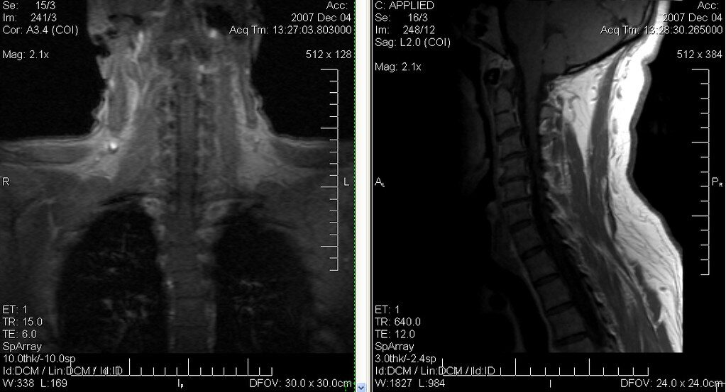
An MRI does not measure pain. It creates detailed images of the structures inside your spine — the vertebrae, intervertebral discs, spinal cord, nerve roots, and surrounding soft tissue. It shows anatomy: what the structures look like at the moment of the scan. What it cannot show is how much pain you are in, how long your symptoms will last, or what your recovery will look like. That context comes from a clinical examination, not from the report alone.
This is the most important thing I tell patients before we discuss anything else on their report: an MRI finding describes a structure. It does not predict your outcome.
What does “disc bulge” or “disc herniation” mean?
A disc bulge means the outer wall of your intervertebral disc has pushed outward slightly beyond its normal boundary. A disc herniation — what many patients call a “slip disc” — means the inner material of the disc has pushed through a tear in that outer wall. Both can press against nearby nerve roots, which is what causes the radiating pain, numbness, or tingling that typically runs into the leg or arm.
It is worth knowing that disc bulges are extremely common — they are found on MRI scans of people with no pain at all. The clinical question is not simply whether a bulge exists, but whether it is causing the symptoms you are experiencing. That distinction changes the entire conversation about treatment.
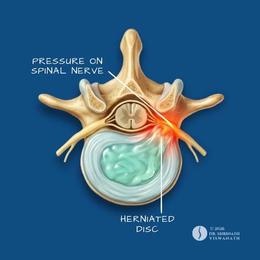
A herniated disc — commonly called a slip disc — occurs when the inner disc material pushes through its outer wall and presses on nearby nerve roots.
What is “foraminal narrowing” or “spinal stenosis”?
The foramen is the small opening on either side of each vertebra through which your nerve roots exit the spinal canal. Foraminal narrowing means this opening has become smaller — usually due to a disc bulge, bone spur, or age-related thickening of surrounding tissue — reducing the space available for the nerve. Spinal stenosis is a broader narrowing of the spinal canal itself.
Neither term automatically means surgery. Many patients with moderate foraminal narrowing manage well with conservative treatment. The severity of your symptoms, not just the appearance of the scan, guides the treatment decision.
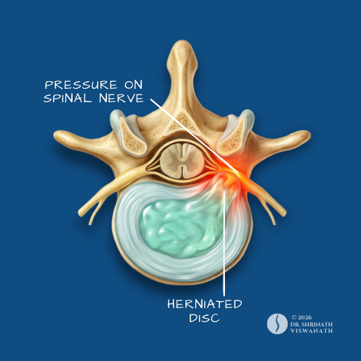
Spinal stenosis refers to a narrowing of the spinal canal. Severity on an MRI image does not always correspond directly to the level of pain a patient experiences.
What does “degenerative changes” mean?
Degenerative changes is a broad term for the natural wear that occurs in the spine over time — reduced disc height, small bone spurs forming at the vertebral edges, changes in the disc itself. These findings are present on the MRI scans of most adults over 40 and are considered a normal part of ageing.
Seeing “degenerative changes” on your report does not mean your spine is damaged beyond help. It means your spine has aged, as all spines do. In my clinic, I am far more interested in what you are feeling day-to-day than in what a scan label says about the age of your disc tissue.
Why does my report say “mild,” “moderate,” or “severe”?
Radiologists use these grades to describe what they see in the images. Mild, moderate, and severe refer to the degree of structural change — how much the disc is pressing on a nerve, how narrow the canal has become. They are useful descriptors, but they are not a treatment prescription.
A “severe” finding on an MRI does not automatically mean severe symptoms, just as a “mild” finding can sometimes cause significant pain if it is pressing precisely on a sensitive nerve root. I always read an MRI report alongside a patient’s clinical history — the two together tell a far more complete story than either one alone.
Can my MRI look serious but my symptoms be mild — or vice versa?
Yes — and this surprises many patients. The correlation between MRI findings and symptom severity is genuinely imperfect. Research consistently shows that significant structural findings on MRI scans — including disc herniations and spinal stenosis — are present in a meaningful proportion of people who have no back pain at all.
The reverse is also true. Some patients with severe, debilitating pain have MRI findings that look relatively unremarkable. This is one of the reasons a thorough physical examination — testing reflexes, muscle strength, sensation — matters as much as, sometimes more than, the scan itself.
When should you see a spine doctor about your MRI findings?
If you have received a spine MRI report, I would recommend a consultation if any of the following apply:
— You have pain, numbness, or weakness that runs into your leg or arm — not just your back or neck.
— You are experiencing weakness in your foot, hand, or leg that is getting progressively worse.
— You have any change in bladder or bowel control — this requires urgent assessment, not a scheduled appointment.
— Your pain is waking you from sleep at night regularly.
— You have been managing with medication or physiotherapy for more than six to eight weeks without meaningful improvement.
— Your report mentions spinal cord compression, cauda equina involvement, or significant instability — these warrant prompt specialist review.
An MRI report is a starting point, not a conclusion. What the scan shows needs to be interpreted in the context of your symptoms, your examination findings, and your daily function — only then does a treatment path become clear.

Patients often ask me: what does your spine MRI report mean for my treatment? The honest answer is that most findings have context, most have options, and most conditions are manageable when approached with accurate information.
If your report has raised questions you cannot answer alone, that is exactly what a consultation is for — not to alarm you further, but to give you a clear picture of where you stand.
To book a consultation, WhatsApp +91-70192-72711 or visit drshrinathspine.com/book-consultation